Radiographic assessments for the early stage of ungular cartilage ossifications (side bone) in donkeys (Equus asinus)
Authors: Latifa A. Alhusayni, Adel I. Almubarak, Zakriya A. Al-Mohamad, Tarik N. Misk, and Sayed F. El-Hawari
Ger. J. Vet. Res
2025.
vol. 5, Iss. 2
pp:24-29
Doi: https://doi.org/10.51585/gjvr.2025.2.0131
Abstract:
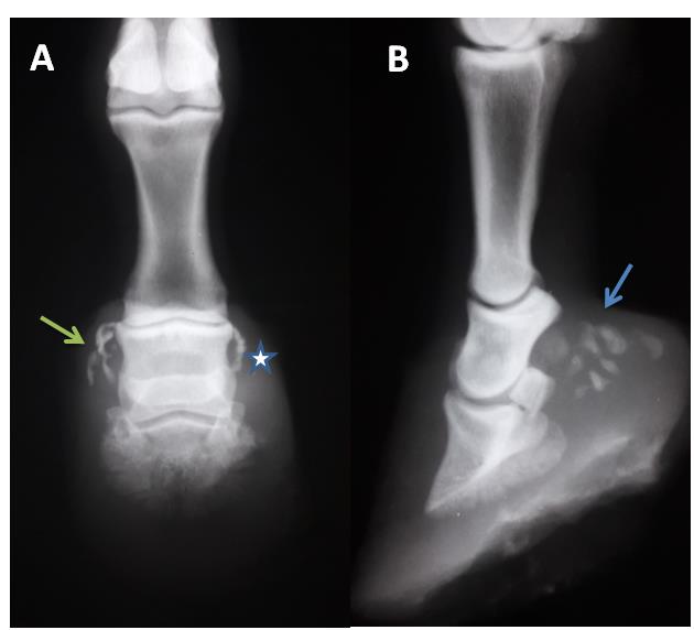
Lameness is a common problem that significantly affects equine health and disrupts overall quality of life. In rural communities, donkeys primarily serve as working animals, and many are afflicted with ossification of the ungular cartilage, commonly known as side bone. This study aimed to provide a radiographic description of an early stage of ungular cartilage ossification (side bone) with a full investigation of accompanying anatomic changes, consequent complications, and adjuvant diseases. A total of 122 radiographic films from 71 donkeys were analyzed. Animal history and lameness degree were then recorded. Standard latero-medial and dorso-palmar radiographic views were reviewed independently by two observers. Side bones were evaluated for their location within the same limb, shape, abnormal radiographic findings, and adjuvant complications. Results revealed the occurrence of ossifications in both right and left thoracic limbs (40.2% and 59.8%, respectively). Bilateral collateral cartilage ossifications in the right (71.5%) and left (79.5%) forelimbs were recorded. Side bone was seen as a solitary irregular ossification center in 29.2% of examined films and appeared as multiple irregular ossification centers in 70.8% of radiographic films. Ossifications appeared in dorso-palmar radiographic films as crescent-like shape radio-opaque structures at the level of the middle phalanx. One area of osteolysis in the pedal bone and widening in the solar foramen were detected in 16.6% of examined radiographic films. In conclusion, the early stage of the side bone occurred bilaterally in donkeys with more extensive ossifications in the lateral cartilage than the medial cartilage. This condition may be associated with lameness and often develops as a sequela to quittor, potentially progressing to septic pedal osteitis. Unlike horses, side bones in donkeys start proximally and extend distally.
Keywords:
Donkey, Radiography, Side bone, Ungular cartilage
Statistics:
Article Views: 113
PDF Download: 7